Prof. Michael Mimouni completed his fellowship at Canada’s leading centre for corneal transplantation. During that period he trained in and performed corneal transplants of every type — full-thickness grafts (PKP), deep anterior lamellar grafts (DALK) and partial-thickness posterior grafts (DMEK and DSAEK) — and gained experience with advanced grafts such as the Boston KPro and with Descemet stripping without any graft at all (DSO).
Prof. Mimouni also maintains a dedicated site with expanded, in-depth information on the subject — corneal transplant — covering the types of transplant, the course of recovery and illustrative cases.
What a corneal transplant is and when it is needed
The cornea is the clear window at the front of the eye. It is not merely a cover but also the most powerful lens in the visual system, so any damage to its clarity or its shape translates directly into loss of vision. A corneal transplant is surgery in which damaged or diseased corneal tissue is replaced with donated tissue — the graft. The tissue is taken from a recently deceased person with no known diseases or other factors that could affect the survival of the tissue or the health of the recipient.
It is important to understand that a transplant is not the first treatment in the chain but the last. We turn to it when vision has been lost because of a structural problem in the cornea that cannot be corrected any other way. The common reasons are advanced keratoconus, in which the cornea has become so distorted that vision can no longer be corrected optically; Fuchs dystrophy and other conditions in which the inner cell layer of the cornea stops functioning and the cornea absorbs fluid and clouds over; a cornea that has lost its clarity following infection, a chemical burn or trauma; and a previous graft that has run its course. Another reason, more common than people tend to think, is weakening of the inner cell layer following cataract surgery, particularly in an eye that was already borderline beforehand.
When a transplant can be avoided
Before discussing a transplant it is worth making sure that the options which do not require donor tissue have been exhausted, because a substantial proportion of the patients who arrive with a referral for a transplant turn out not to need one.
In keratoconus, cross-linking performed in time halts the progression and usually makes a transplant unnecessary altogether. Even when the cornea has already become significantly distorted, rigid or scleral contact lenses often restore good vision without any surgery, simply because the lens creates a new optical surface above the irregular cornea. In the early stages of Fuchs dystrophy it is sometimes possible to strip only the diseased Descemet membrane and allow the remaining healthy cells to repopulate the area — a procedure known as DSO, which involves no tissue transplantation at all. And in eyes where the central problem is an inflamed ocular surface, treating that surface properly before any decision can change the whole picture.
The decision rests on measurement rather than impression. Corneal topography shows the shape and thickness at every point, and complementary tests count the endothelial cells and measure the degree of swelling. It is the combination of the two that determines whether a transplant is needed, which type is appropriate, and whether it is still reasonable to wait.
Partial or full-thickness — the principle that changes everything
The cornea is not a uniform tissue. It is only about half a millimetre thick (around 550 microns), and it is built in layers: an outer epithelium, a thick stroma that makes up most of the thickness, and, on the inside, Descemet membrane and the endothelial cell layer, which pumps fluid out and keeps the cornea dry and clear.
The principle that guides modern corneal surgery is simple: replace only the diseased layer. For decades there was one option alone — replacing the full thickness of the cornea — and every patient received the same operation regardless of which layer had failed. Today the situation is different. When the problem lies in the front layers, we replace those and leave the patient their own endothelial cells. When the problem lies in the inner layer, we replace that alone — sometimes less than five per cent of the corneal thickness — and leave the rest of the cornea in place. Only when all the layers are damaged do we replace all of them.
This division has three practical consequences: the less tissue replaced, the lower the risk of graft rejection, the faster the recovery, and the stronger the eye remains structurally, because there is no full-depth circumferential incision. The four established types — PKP, DALK, DMEK and DSAEK — differ from one another on exactly this question: which layer is replaced and which stays.
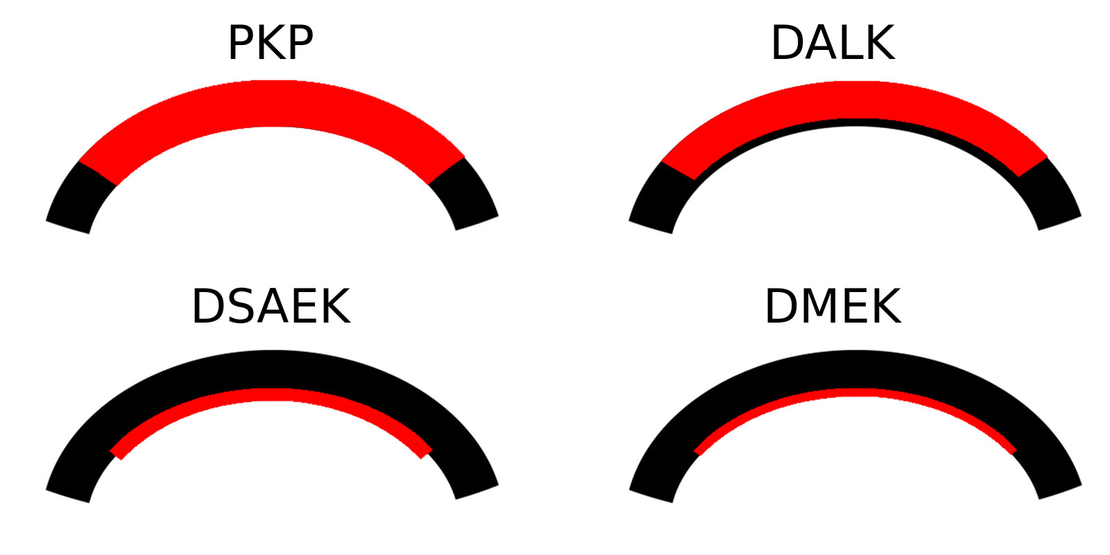
Full-thickness corneal transplantation (PKP)
A full-thickness transplant, known as PKP (short for Penetrating Keratoplasty), is performed when all the layers of the cornea are damaged and neither an anterior nor a posterior lamellar graft is possible. This situation is typical of deep scarring after infection or trauma, of very advanced keratoconus with central scarring, and of previous grafts that have failed.
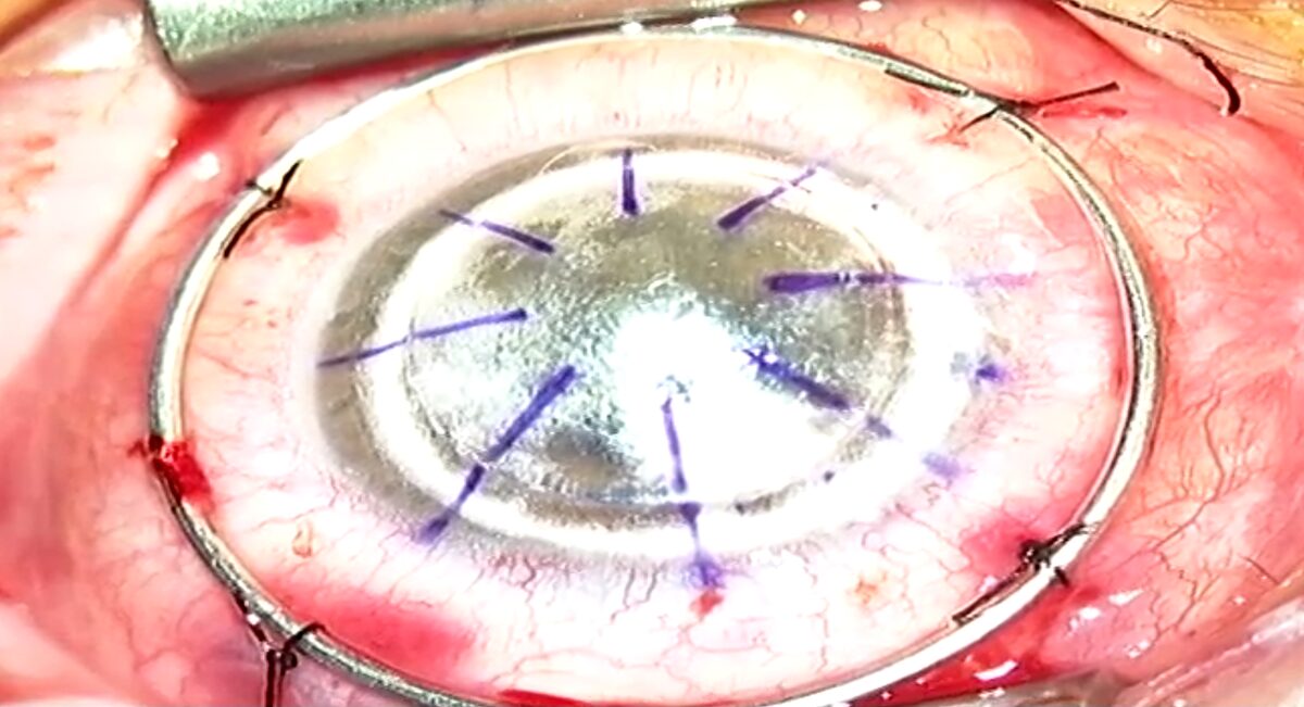
In the operation a full-thickness disc is removed from the patient’s cornea and replaced with a disc of matching diameter from the donor cornea. The graft is sutured into place with extremely fine sutures, thinner than a hair. It can be secured with a single continuous suture running around the entire circumference, or with a series of individual sutures — usually about sixteen — each tied separately. The advantage of individual sutures is that they can be removed selectively later on in order to correct astigmatism, so they are used when fine-tuning of the vision is expected to be needed. During surgery a special metal ring is sometimes sutured to the wall of the eye to preserve the structural strength of the globe at the moment when the cornea is open — the most delicate moment of the procedure.
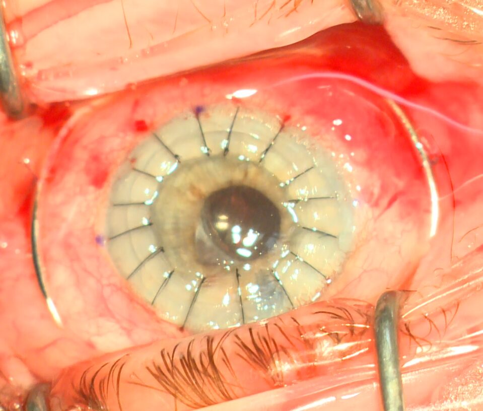
Recovery from a full-thickness graft is the longest of all the types. We wait about eight to twelve months before beginning to remove sutures, in order to allow the tissue to heal and integrate properly, and vision keeps changing for as long as the sutures are in place. At the end of the process most patients need glasses or a contact lens to get the most out of the graft, because suturing almost always leaves some degree of astigmatism.
Deep anterior lamellar keratoplasty (DALK)
A deep anterior lamellar transplant, known as DALK (short for Deep Anterior Lamellar Keratoplasty), is performed when the front layers of the cornea are damaged — in keratoconus, for example — but the endothelial cell layer is healthy. In that case there is no reason at all to replace the whole cornea, and only the stroma above Descemet membrane is replaced.
The main advantage is that the patient’s own natural endothelial cells are preserved. This matters because rejection of a corneal graft is in most cases directed precisely against the donor’s endothelial cells — and when those cells do not come from a donor, that risk all but disappears. In addition, because the eye is not opened to its full depth, it remains structurally stronger.
The technical challenge is separating the stroma from Descemet membrane, which is only a few microns thick. The technique used today is based on injecting a large air bubble into the thickness of the cornea, which separates the layers from one another along a natural plane far more precisely than any manual dissection could. The method carries the name of Mohammed Anwar, who described it, and it is considered the standard because it produces better results than manual peeling. Here too the graft relies on sutures, and here too recovery is measured in months — but it is somewhat faster than after a full-thickness graft, and suture removal can begin earlier.
Posterior grafts: DMEK and DSAEK
In the last two techniques the problem is the reverse: the front layers of the cornea are intact, and it is the inner cell layer that is damaged or not functioning. This is the situation in Fuchs dystrophy and also in the corneal clouding that sometimes develops after intraocular surgery. In both cases the cornea absorbs fluid, swells and loses clarity, and vision is blurriest in the morning hours.
Grafts of this kind have transformed the field from end to end. They do not require opening the eye to its full depth; instead a thin sheet of tissue is introduced through a small incision and pressed against the cornea from the inside using a bubble of air or gas. It is that bubble which holds the graft in place during the first days, which is why the patient is asked to lie on their back for many hours after surgery — an instruction that sounds trivial but is one of the strongest determinants of whether the operation succeeds.
DMEK
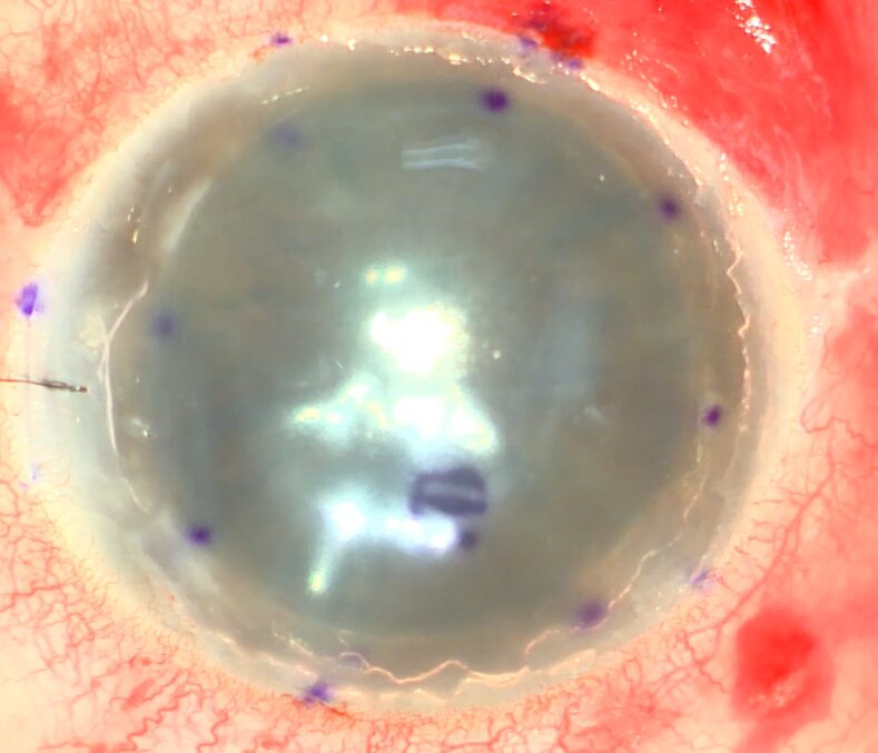
In DMEK (short for Descemet Membrane Endothelial Keratoplasty) only Descemet membrane and the cell layer attached to it are replaced. The graft is less than twenty microns thick, meaning less than five per cent of the corneal thickness. It is the most delicate of all the grafts: it arrives rolled up like a tiny scroll and has to be unfolded inside the eye and confirmed not to be upside down, and for that purpose a letter is marked on the tissue which allows the correct orientation to be identified and which fades away on its own within a few days.
In exchange for that delicacy comes the best result available: because no foreign stroma is added, vision is sharper, recovery is measured in weeks rather than months, and the risk of rejection is lower than with any other graft. The graft usually needs no sutures at all, or at most one or two to close the incision. In about ten to twenty per cent of cases the graft does not attach completely and an additional air bubble has to be injected in the first weeks. That sounds like a complication, but it is a short procedure in the clinic, and it is an anticipated part of the path rather than a failure.
DSAEK
In DSAEK (short for Descemet Stripping Automated Endothelial Keratoplasty) the inner cell layer is replaced together with a thin layer of stroma carrying it, so the graft thickness ranges from sixty to a hundred and fifty microns depending on the cutting capability of the instrument. The thicker graft is stiffer, and it is far easier to unfold and position inside the eye.
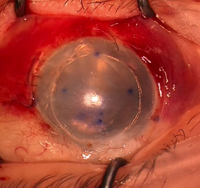
DSAEK therefore remains the preferred technique in situations where DMEK is not possible or would be too complex: poor visibility into the eye, very long eyes, eyes that have undergone vitrectomy, and other situations in which controlling an extremely thin sheet of tissue is difficult. The graft requires a few individual sutures to close the incision, which can be removed as early as a month after surgery. Recovery is measured in months rather than weeks, and the final vision is good but usually slightly less sharp than after DMEK, because a layer of foreign tissue has been added within the optical axis. Here too an additional air injection may be needed, though less often than after DMEK.
What recovery looks like
In the first days vision is blurred, the eye is sensitive to light, and watering and a foreign-body sensation are possible. After posterior grafts patients are asked to lie on their back so that the bubble presses the graft into place, and after sutured grafts the main restriction is avoiding anything that could put pressure on the eye: rubbing, lifting heavy loads, prolonged bending and contact sports.
Medical treatment is based on steroid drops, which reduce inflammation and prevent rejection, and on antibiotic drops in the first weeks to prevent infection. The steroids are the part that demands persistence: they are given frequently at first and tapered very slowly over months, and some patients continue on a low dose for years. Stopping the drops independently is one of the most common causes of a graft rejection that would otherwise have been avoided.
The timetable depends on the type of graft: after DMEK most patients see a marked improvement within weeks, after DSAEK within months, and after PKP and DALK vision keeps changing for as long as the sutures are in place and settles only after one to two years. Alongside this, dry eye is a common companion of this period — the corneal nerves are cut during surgery and recover slowly, and in the meantime the burning sensation and the fluctuating blur can be more troubling than the graft itself. Preservative-free lubricating drops are a fixed part of the treatment.
Graft rejection — the signs that require urgent attention
Rejection does not mean the graft is lost. When it is identified early it can usually be halted with intensified steroid treatment, and the later it is identified the greater the irreversible damage to the endothelial cells. Four signs call for attention the same day, even years after surgery: new redness in the eye, increased sensitivity to light, a drop in vision, and pain. None of them is part of normal recovery at a late stage, and a common mistake is to attribute them to tiredness or dryness and wait for the next scheduled appointment.
What the public health basket covers
Unlike elective procedures, a corneal transplant is medically necessary surgery, and it is included in the public health basket in Israel — donor tissue included. The tissue comes from eye banks and is managed through the national system, and when local supply is insufficient tissue is imported from banks abroad. Follow-up and the drops used afterwards also fall within the health fund’s regular service.
What does differ between patients is the route: surgery within the public system at the hospital to which the patient was referred, as opposed to choosing your surgeon through the fund’s supplementary insurance or through private insurance. The terms of participation and eligibility differ between funds and between levels of cover, and they are updated from time to time, so it is worth checking with your fund what is currently in force before fixing a date.
One further point that many people are unaware of: when vision remains impaired over time, whether before the transplant or after it, there may be an entitlement to a disability rating for visual impairment. I have built a disability rating calculator for visual impairment that lets you check the initial picture before approaching a committee.
Evaluation and consultation with a cornea specialist
Whether a corneal transplant is needed, and which type is appropriate, is not answered by the diagnosis alone. Two patients with the same disease can need entirely different operations depending on which layer has failed, on the state of the endothelial cells, on the shape of the cornea and on the condition of the ocular surface. Prof. Michael Mimouni is a specialist in cornea and ocular surface, completed his fellowship at Canada’s leading centre for corneal transplantation, and performs all types of graft — including the delicate posterior techniques.
An examination establishes what exactly is causing the loss of vision, whether a transplant can still be avoided, and if not, which type suits this particular eye and what it is realistic to expect from it. Further information on other conditions that can affect vision can be found on the eye diseases page. To arrange an examination, for a second opinion or with any questions, you are welcome to get in touch. When and why to see a doctor who specialises in the cornea is explained in cornea specialist.
Frequently asked questions
How long does it take to recover from a corneal transplant?
It depends on the type of graft. After a DMEK graft most patients see a significant improvement within a few weeks, and after DSAEK within a few months. Recovery from a full-thickness graft is the longest: vision keeps changing for as long as the sutures are in place, so this is a process of one to two years until it fully stabilises.
Is a corneal transplant painful?
The surgery itself is not painful — it is performed under local or general anaesthesia. In the first days afterwards a foreign-body sensation, watering and light sensitivity are common, but significant pain is not typical. New pain appearing weeks or months after surgery is not part of normal recovery and needs to be examined.
What are the chances the graft will take?
In low-risk corneal transplants success rates are high, and most grafts are clear and functioning at the end of the first year. The odds depend mainly on the reason the transplant was needed: a quiet eye with little inflammation and no blood vessels in the cornea is the best-case scenario, while an eye with scarring after infection, vessels that have grown into the cornea, or a previous failed graft carries a higher risk.
How long does a corneal graft last?
A corneal graft is not necessarily for life. Some grafts function for decades, while in other patients the endothelial cells are gradually depleted over the years and the graft loses its clarity. When that happens a repeat transplant is usually possible, and it is often only a partial posterior graft rather than a replacement of the whole cornea.
Is there a long wait for donor corneal tissue?
Corneal tissue comes from eye banks and is managed through the national system, and when local supply is insufficient tissue is imported from banks abroad. In practice corneal transplants do not work like urgent organ transplants: in most cases this is planned surgery scheduled in advance, not a wait for a phone call.
Is a corneal transplant covered by the public health basket in Israel?
Yes. Unlike elective procedures such as laser vision correction, a corneal transplant is medically necessary surgery and is included in the public health basket, tissue included. What can differ between patients is the route — surgery within a public hospital as opposed to choosing your surgeon through supplementary or private insurance. Terms vary between health funds and are updated from time to time.