Pterygium – Understanding Its Causes, Risk Factors and Treatments
Definition
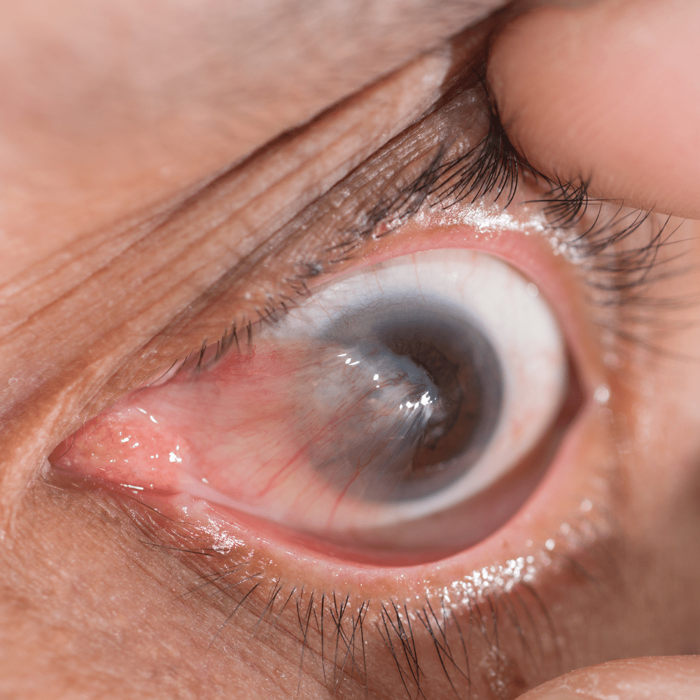
Pterygium, often referred to as “surfer’s eye”, is a common eye condition in which an abnormal, non-cancerous growth develops on the conjunctiva – the thin, transparent layer of tissue covering the white of the eye – and extends onto the cornea, the clear front surface of the eye. This benign growth typically takes on a wing-like or triangular shape, which gives it its name, derived from the Greek word “pterygion”, meaning “wing”.
In its early stages, a pterygium may be small and barely noticeable, causing little or no discomfort. As it grows, however, it can become a significant problem, potentially distorting vision, causing discomfort, or raising concerns about appearance. Some people may notice a foreign-body sensation, itching or redness associated with the growth. A larger or more aggressive pterygium can induce astigmatism by altering the shape of the cornea, or can encroach on the visual axis, leading to impaired vision. It is important to note that while a pterygium is usually benign, its presence – particularly when symptomatic – warrants evaluation by an ophthalmologist to determine appropriate treatment.
Causes of Pterygium
The exact cause of pterygium is not fully understood, but the prevailing view is that the condition results from a combination of genetic predisposition and environmental factors. The leading theory points to prolonged exposure to ultraviolet (UV) light, most often from the sun, as a significant risk factor. UV light can damage the DNA within the cells of the eye, triggering an inflammatory response that stimulates fibrovascular growth, ultimately leading to the formation of a pterygium.
In addition to UV exposure, other environmental elements such as dust and wind are also considered significant contributors. These elements can cause microtrauma to the surface of the eye, leading to cellular changes in the conjunctiva and cornea and promoting the growth of a pterygium. Furthermore, ocular surface disease has been linked to the development of pterygium. It is thought that a deficiency in the protective tear film may leave the eye vulnerable to the harmful effects of UV radiation, dust and wind.
Finally, certain biochemical factors may also play a role. For example, growth factors and cytokines – proteins involved in cell signaling – are believed to be involved in the development and progression of pterygium. These substances can promote cell proliferation, migration and matrix remodeling, the key features of pterygium growth. In particular, overexpression of vascular endothelial growth factor (VEGF) and matrix metalloproteinases (MMPs) has been identified in pterygium tissue.
While these factors offer some insight into the possible causes of pterygium, further research is needed to fully clarify the underlying pathophysiological mechanisms. It is essential for individuals, especially those in high-risk groups, to take preventive measures such as wearing UV-protective glasses when exposed to intense sunlight or harsh environmental conditions.
Pterygium – Risk Factors
The development of a pterygium is associated with several risk factors, many of which relate to both geographic location and lifestyle choices.
Ultraviolet radiation: The most significant risk factor for pterygium is prolonged exposure to ultraviolet (UV) radiation. The outer surface of the eye is directly exposed to sunlight, and chronic exposure can promote the growth of a pterygium. UV radiation has been found to damage limbal stem cells, which can lead to uncontrolled proliferation of conjunctival tissue onto the cornea.
Age: Pterygium primarily affects adults, with prevalence increasing from age 30 onward. The risk rises with age as the cumulative effect of UV radiation and environmental exposure contributes to the development of the condition.
Geography: Geographic location plays a crucial role in the prevalence of pterygium. It is more common among people living closer to the equator, an area often referred to as the “pterygium belt”, where the sun’s rays are strongest.
Outdoor activities: Occupations or hobbies that involve prolonged outdoor exposure increase the risk of pterygium. Outdoor workers such as farmers, fishermen and construction workers are at higher risk. Similarly, those who take part in outdoor sports or recreational activities without adequate eye protection are also more exposed.
Sex: Some studies suggest that men may be slightly more prone to pterygium than women, likely due to increased outdoor activity and occupational (sun) exposure.
Pterygium – Treatments
The approach to treating a pterygium is individualized, taking into account its size, location, rate of growth and the extent of symptoms.
Observation only: For a small, asymptomatic pterygium, a “watch and wait” approach may be adopted. Regular eye examinations can ensure that any change in size or the onset of symptoms is addressed promptly.
Eye drops and ointments: If the pterygium causes discomfort or redness, lubricating eye drops or artificial tears can be used to relieve dryness and irritation. For a more inflamed or aggressive pterygium, topical steroid eye drops can help reduce inflammation and soothe the redness of the eye.
Contact lenses: Specialized therapeutic contact lenses can provide a physical barrier against wind and dust, protecting the pterygium from further irritation.
Sunglasses: Wearing sunglasses that block UV light can help slow the growth of a pterygium, especially for those frequently exposed to sunlight.
Natural Treatment for Pterygium — What Actually Helps and What Does Not
Many people search for a “natural treatment” for pterygium and come across all sorts of promises. It is important to say this honestly: there is no natural remedy, drop or medication that makes an existing pterygium disappear. The tissue that has grown over the conjunctiva does not “retreat” on its own. What conservative measures can do is relieve the symptoms and slow the progression:
- Protection from sun and wind — pterygium is strongly linked to cumulative UV exposure. Quality sunglasses with UV protection and a peaked hat are the only thing that genuinely reduces the factor driving the growth, and therefore also the most effective “natural” measure.
- Artificial tears — they do not shrink the pterygium, but they relieve the burning, redness and gritty sensation that arise from dryness and irritation over the growth.
- Reducing irritation — avoiding dust, smoke and dry wind, and sometimes periodic drops to calm local inflammation as needed.
The conservative approach is appropriate when the pterygium is small, stable and does not interfere with vision. But it manages the condition — it does not remove it. When the pterygium grows, causes persistent discomfort, becomes red again and again, or approaches the centre of the cornea and threatens vision, the only way to remove it is surgery. So anyone promising to “get rid of a pterygium without surgery” is not being accurate.
How Much Does Pterygium Surgery Cost
The cost of pterygium surgery varies according to several factors: whether it is a primary or a recurrent pterygium (repeat surgery is more complex), the technique chosen (particularly the use of a conjunctival autograft or an amniotic membrane to reduce recurrence), and the setting in which the surgery is performed. A private procedure, a procedure through supplementary insurance, or an arrangement with a health fund — each is priced differently.
Beyond the price itself, it is worth knowing: pterygium surgery performed with the right technique, with an autograft rather than a simple “scraping,” is more expensive but significantly reduces the chance of recurrence — and that saves a further operation later on. What is cheap in the short term is not necessarily cheap in the long term.
Recovery After Pterygium Surgery
The surgery itself is short and is performed under local anaesthesia. In the first days afterwards the eye is red and sensitive, with a foreign-body sensation and watering — this is expected and stems from the healing of the ocular surface. You feel gradual improvement over the first week or two, and the redness fades over several weeks until the eye looks white again. During this period it is important to use the drops that were prescribed, to avoid rubbing the eye, and to be diligent about sun protection — precisely the factor that contributed to the pterygium in the first place.
Most people return to their routine within a few days, but full healing of the surface takes time, and adhering to the instructions during this period is what reduces the chance of recurrence. Some patients have a tendency to recurrence, and so follow-up after the surgery is part of the treatment.
Indications for Pterygium Surgery
Surgical intervention becomes a consideration under several circumstances:
Persistent discomfort: If the pterygium causes chronic irritation, redness or a foreign-body sensation that is not relieved by eye drops or artificial tears, surgery may be recommended.
Visual impairment: When the growth encroaches on the central area of the cornea or causes significant astigmatism due to its size and location, it can impair visual acuity. In such cases, surgical removal is often recommended.
Rapid progression: A rapidly growing pterygium can pose a threat to vision and requires surgical intervention to prevent further ocular complications.
Cosmetic reasons: A large or inflamed pterygium can be quite conspicuous, causing distress for some people because of its appearance. If the appearance of the pterygium significantly affects a person’s quality of life, surgical removal may be considered.
Failure of medical treatment: If pterygium symptoms do not improve, or worsen, despite non-surgical treatments, surgery may be the next step.
In all cases, the decision to operate should be made after a detailed discussion between the patient and the ophthalmologist, weighing the benefits of surgery against potential complications.
Surgical Techniques
Surgical techniques for pterygium have evolved over the years to improve outcomes and reduce recurrence rates.
Excision with autologous graft: This technique remains the most common surgery for pterygium. It involves careful removal of the pterygium followed by transplantation of conjunctival tissue, often taken from beneath the patient’s eyelid, to the site from which the pterygium was removed. The graft, usually secured with sutures or fibrin glue, helps fill the defect and prevent recurrence by inhibiting regrowth of pterygium tissue.
Excision with amniotic membrane grafting: In this procedure, after excision of the pterygium, an amniotic membrane graft is used to cover the defect. The amniotic membrane, which is the innermost layer of the placenta, has anti-inflammatory, anti-scarring and healing-promoting properties that can enhance recovery and reduce recurrence.
Conjunctival autograft with fibrin glue: This technique is a modification of the traditional autograft procedure. Instead of sutures, fibrin glue is used to attach the conjunctival graft. The glue mimics the final stages of the body’s clotting mechanism, creating a firm bond between the graft and the scleral tissue. The use of fibrin glue may reduce operating time and postoperative discomfort while providing excellent graft stability.
Application of mitomycin C: Mitomycin C, an antiproliferative agent, is sometimes applied during surgery to further reduce the likelihood of pterygium recurrence. It inhibits fibroblast proliferation, thereby reducing scar formation and regrowth of pterygium tissue.
Complications of Pterygium Surgery
Despite ongoing advances in surgical techniques, pterygium surgery, like any procedure, can be associated with several complications:
Recurrence: This is the most common complication following pterygium surgery. The recurrence rate can be as high as 40% with simple excision but drops significantly with conjunctival membrane grafting or amniotic membrane grafting.
Infection: Although rare due to the routine use of prophylactic antibiotics, infections can occur after surgery. Patients should be aware of symptoms such as increased redness, pain, discharge or a sudden decrease in vision, which may indicate an infection.
Graft complications: These can include displacement, retraction or rejection of the graft. Although rare with autografts, they can occur, particularly when amniotic membrane grafts are used.
Scleral thinning or perforation: These are rare but serious complications that can occur, particularly during repeat surgeries or extensive procedures in which mitomycin C is used excessively.
Astigmatism: Surgery can alter the shape of the cornea, leading to astigmatism, which may affect visual acuity. This is usually temporary and diminishes as the eye heals.
Dry eye syndrome: Some patients may experience dryness of the eye after surgery, which can usually be managed with artificial tears or ointments.
Symblepharon formation: This involves adhesion of the eyelid to the eyeball, a rare complication usually associated with extensive surgeries.
Corneal scarring or opacities: In rare cases, corneal scarring can occur following surgery, which may affect vision.
The potential for complications underscores the importance of thorough preoperative evaluation, meticulous surgical technique and close postoperative follow-up. Patient education is essential to ensure an understanding of potential risks and adherence to postoperative care instructions in order to improve surgical outcomes.