If you have been referred for “corneal mapping” — before laser surgery, following a spectacle prescription that keeps changing, or as part of an investigation — it is worth understanding what this examination looks at and why it is so central to corneal medicine. It is a simple, non-invasive examination, but the information it provides is often what determines the diagnosis and the right treatment.
What Corneal Mapping Is
The cornea is the transparent “window” at the front of the eye, and its precise shape is what determines how light is focused. Corneal mapping — topography, and on advanced devices tomography as well — creates a detailed map of the surface of the cornea and of its thickness, much like a topographic map of mountains and valleys. The examination measures the curvature at every point and identifies tiny irregularities in shape that cannot be seen in a routine eye examination.
The examination itself takes minutes, involves no contact with the eye and is not painful: you look into a device that scans the cornea, and that is all.
When Corneal Mapping Is Required
The examination is used in several key situations:
- Diagnosing and monitoring keratoconus. Corneal mapping is the principal tool for identifying a conical cornea — sometimes before there are clear symptoms — and for monitoring whether the condition is stable or progressing. It is also the examination that determines whether cross-linking treatment is needed.
- Before laser vision correction. Every candidate for laser surgery must have preliminary corneal mapping. The examination confirms that the cornea is regular and thick enough, and above all rules out latent keratoconus, in which laser surgery is contraindicated. This is one of the most important screening steps for the safety of the surgery.
- Planning and assessment in a diseased cornea. In situations such as scarring, irregularity following previous surgery, or assessment ahead of a corneal transplant, the map helps in understanding the state of the cornea and planning the treatment.
- Investigating vision that cannot be corrected. When glasses do not give sharp vision, or when the prescription changes again and again, corneal mapping helps establish whether the cause lies in the shape of the cornea.
What the Examination Reveals
The map shows the curvature pattern of the cornea and the areas of steepening or thinning. Certain patterns are characteristic of keratoconus; others indicate regular astigmatism that can be corrected with glasses; and others an irregularity that requires special contact lenses. Thanks to the map it is possible to distinguish between conditions that look similar on a routine examination but require entirely different treatment — and that is exactly why it is such a central tool.
How to Read Corneal Mapping Results
Many patients receive the mapping printout and try to make sense of it on their own. The explanation below will help you find your way around it, but the interpretation itself is always made together with the eye examination, not from the printout alone.
The Main Maps on the Printout
Most devices display several maps side by side:
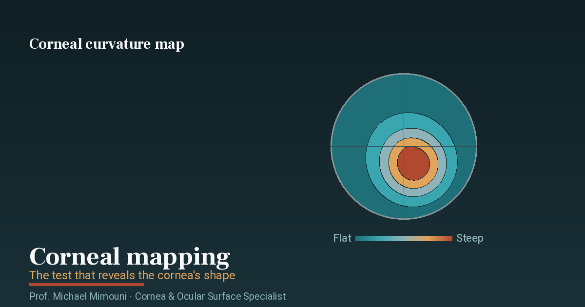
- The curvature map (axial or sagittal) shows how steep or flat the cornea is at each point. This is the most familiar map.
- The elevation maps, front and back, compare the corneal surface with an average spherical shape and show areas that rise above it or dip below it. The back elevation map is particularly sensitive to early changes of keratoconus.
- The thickness map (pachymetry) shows the thickness of the cornea at each point, and above all where the thinnest point is and how thick it is.
What the Colours Mean
On the curvature map, warm colours (red and orange) mark steeper areas, and cool colours (blue and green) mark flatter areas. On the other maps colour has a different meaning, so always check the map’s title and the colour scale beside it. The scale itself can also vary: when it is set to a narrow range, bright red can represent a very small difference. So “a lot of red” is not necessarily a problem.
The Important Numbers
- K1 and K2 are the curvature of the flat axis and of the steep axis, in dioptres. The difference between them reflects the astigmatism of the cornea, and the axis shows its direction.
- Kmax is the steepest point of the cornea. In keratoconus it is one of the values followed over time.
- The thickness at the thinnest point, and its location. In a normal cornea the thinnest point is close to the centre. A thinnest point displaced downwards or to the side is one of the signs that are checked.
- Keratoconus screening indices. Many devices calculate combined indices and mark them in colour: white or green for normal, yellow for borderline, red for abnormal. This is an important aid, but not a diagnosis. A yellow value can also result from dry eye or an imprecise measurement, and a normal value does not replace a full examination when there is suspicion.
Patterns the Doctor Looks For
- A symmetric “bow tie” indicates regular astigmatism, which is corrected well with glasses or contact lenses.
- An asymmetric bow tie, or two halves that are not on the same line, is one of the signs that raise suspicion of keratoconus, especially when there is also steepening in the lower part of the cornea.
- A localised steep area, usually below the centre, together with thinning in the same area, is a picture characteristic of keratoconus.
- A flat, even centre after laser surgery for short-sightedness is expected and normal.
- Diffuse irregularity can result from a scar, from previous surgery, or simply from an unstable tear film at the time of the examination.
Comparison Over Time
When monitoring keratoconus, a single map is not enough: what counts is the comparison between examinations. According to the international consensus on keratoconus, progression is defined as a consistent change in at least two of the following: steepening of the front surface of the cornea, steepening of the back surface of the cornea, or thinning of the cornea. For the comparison to be reliable, it is best to perform the examinations on the same type of device, because different devices do not measure in exactly the same way. That is also why you should bring your previous maps to the visit.
What Can Distort the Results
- Contact lenses temporarily change the shape of the cornea. Before mapping, and especially before laser surgery, you are asked to stop wearing them for a set period: usually a few days for soft lenses and longer for rigid lenses. Ask the clinic what the instruction is in your case.
- Dry eye and an unstable tear film can create apparent irregularity on the map. Sometimes the dryness is treated first and the examination is then repeated.
- Blinking, movement or poor fixation of the gaze during the scan. That is why several measurements are usually taken and the best-quality one is chosen.
The bottom line: the printout is one piece of the picture. The decision about whether there is keratoconus, whether it is progressing and whether laser surgery is possible is made only in combination with the eye examination, the medical history and the comparison with previous examinations.
When to See a Cornea Specialist
It is worth having corneal mapping and an evaluation by a cornea and ocular-surface specialist if:
- You are considering laser vision correction — the examination is a prerequisite
- You have been diagnosed with keratoconus, or it is suspected, and you want to know whether the condition is stable or progressing
- Your vision is not fully corrected with glasses, or your prescription changes again and again
- There is a family history of keratoconus
- You have received a mapping printout with values flagged in yellow or red and want to understand what they mean
I perform the examination and its interpretation at my clinic in Haifa, serving patients from across northern Israel. Correct interpretation of the map — not merely performing the examination — is what makes it a genuine diagnostic tool. More on what a visit to a cornea specialist involves is on a separate page.
If you have been referred for corneal mapping, or you want to establish the state of your cornea, you are welcome to get in touch and schedule an examination at my Haifa clinic.
Frequently Asked Questions
Is corneal mapping painful? No. The examination is non-invasive and does not involve contact with the eye. You look into a scanning device for a few minutes, and that is all. No special preparation is required, although contact-lens wearers are sometimes asked to remove them before the examination.
Why is corneal mapping needed before laser surgery? Because the examination confirms that the cornea is suitable for surgery — regular and thick enough — and above all rules out latent keratoconus, a condition in which laser surgery may cause harm. This is one of the most important screening steps for the safety and success of the surgery.
Can corneal mapping diagnose keratoconus? Yes. Corneal mapping is the principal tool for diagnosing and monitoring keratoconus, and it sometimes identifies it at an early stage before clear symptoms appear. It is also the examination on which the decision about whether treatment to stabilise the cornea is needed is based.
How often is the examination repeated? It depends on the reason. When monitoring active keratoconus, the mapping is repeated at regular intervals in order to identify progression; in a stable condition, or as a one-off examination before surgery, routine repetition is not required. The frequency of follow-up is determined according to the individual situation.
What does a red area on a corneal map mean? On the curvature map, red marks a steeper area. That is not necessarily a problem: the meaning depends on the colour scale, the location of the area and its shape. A steep, asymmetric area in the lower part of the cornea, especially together with thinning, raises suspicion of keratoconus, but the diagnosis is made only through a full examination and comparison with previous maps.
Frequently asked questions
Is corneal mapping painful?
No. The examination is non-invasive and does not involve contact with the eye. You look into a scanning device for a few minutes, and that is all. No special preparation is required, although contact-lens wearers are sometimes asked to remove them before the examination.
Why is corneal mapping needed before laser surgery?
Because the examination confirms that the cornea is suitable for surgery — regular and thick enough — and above all rules out latent keratoconus, a condition in which laser surgery may cause harm. This is one of the most important screening steps for the safety and success of the surgery.
Can corneal mapping diagnose keratoconus?
Yes. Corneal mapping is the principal tool for diagnosing and monitoring keratoconus, and it sometimes identifies it at an early stage before clear symptoms appear. It is also the examination on which the decision about whether treatment to stabilise the cornea is needed is based.
How often is the examination repeated?
It depends on the reason. When monitoring active keratoconus, the mapping is repeated at regular intervals in order to identify progression; in a stable condition, or as a one-off examination before surgery, routine repetition is not required. The frequency of follow-up is determined according to the individual situation.
What does a red area on a corneal map mean?
On the curvature map, red marks a steeper area. That is not necessarily a problem: the meaning depends on the colour scale, the location of the area and its shape. A steep, asymmetric area in the lower part of the cornea, especially together with thinning, raises suspicion of keratoconus, but the diagnosis is made only through a full examination and comparison with previous maps.