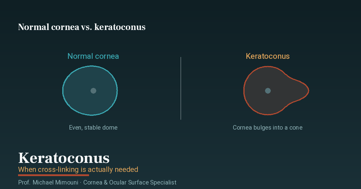
Keratoconus is the most common corneal disease in young patients, and it reaches medical boards again and again — in part because it strikes vision in the years when people are working, studying and serving. And yet anyone searching for the word “keratoconus” in the National Insurance impairment schedule will not find it: it does not appear even once in the ophthalmology chapter. How the disease is nevertheless assessed, and where most of the errors occur, is the subject of this page.
The data on this page comes from the same impairment schedules on which the eye disability percentage calculator is built — the same clauses, the same figures.
There is no keratoconus clause — there is a table
As with cataract and with dry eye, keratoconus is assessed not by diagnosis but by outcome: what the disease has actually done to visual acuity. The visual acuity table intersects the best corrected acuity of the two eyes, and the intersection determines the percentage.
Since keratoconus is usually asymmetric, the typical picture is one eye affected more severely: an eye at 6/60 against a healthy fellow eye — 20%; 3/60 — 25%; a reduction in the fellow eye as well climbs the table quickly. The exact combination of any two acuities appears in the full table on the calculator page.
The decisive question: glasses or lenses
Here lies the heart of the whole keratoconus subject — the point that is almost always missed. The table provides that visual acuity is measured with full optical correction, and the schedule does not specify by what means of correction.
In keratoconus the difference is not theoretical. A conical cornea creates irregular astigmatism that glasses correct only partially, whereas a rigid (RGP) or scleral lens creates a new optical surface and corrects far more: the very same eye may see 6/60 in glasses and 6/9 with a lens. Between those two measurements lie dozens of percentage points of disability in the table.
Proper preparation for a board is therefore not a single measurement but a whole picture: acuity documented in both forms of correction, alongside reliable documentation of actual tolerance — whether the patient can wear the lenses, for how many hours a day, and whether there are medical reasons limiting their use. A file showing only the best measurement, or only the worst, tells half the truth — and in complex files this is precisely where an expert opinion addressing the question head-on makes the difference.
Corneal scarring: clause 61 and its ceiling
Advanced keratoconus can leave a central scar — after acute hydrops, for example. Here clause 61 — corneal opacities comes in: an opacity 2 mm or more in diameter, within the central 4 mm, gives 5% for one eye or 10% for both — but only in the absence of a visual impairment that confers disability. The schedule states it directly: where an opacity causes a reduction in acuity that confers disability, disability is determined by visual acuity alone.
In other words: a small scar that does not affect vision receives the opacity percentage; a scar that reduces vision is absorbed into the table assessment. There is no double track.
After a corneal transplant
When keratoconus reaches the stage requiring a corneal transplant — deep anterior lamellar (DALK) or penetrating (PKP) — the assessment continues to work on the same principle: what decides is not the transplant but the outcome. The best corrected acuity of the grafted eye in a stable state is what goes into the table.
Timing carries particular weight here. After a penetrating graft, roughly eight to twelve months pass before suture removal begins, and vision keeps changing while the sutures are in place; once they are out, astigmatism requiring glasses or a lens usually remains. A measurement taken in the middle of the process does not reflect the final state — in either direction — which is why boards value documentation of a stable state, preferably on more than one date.
Cross-linking and its place in the picture
Cross-linking is intended to halt the progression of the disease rather than to improve vision substantially, so its direct effect on the percentage is limited. Its significance for the board is indirect but real: a series of corneal topographies documenting progression before treatment and stability afterwards is exactly the kind of serial, objective documentation that allows a board to establish findings with confidence.
Weighting, the ceiling, and the Ministry of Defense
Where there are impairments in both eyes or additional defects, the percentages are weighted — each further one taken from the remainder, so 20% plus 10% gives 28%, not 30% — and total disability for one eye is capped at the percentage for a completely blind eye. In general disability it is also worth remembering the exclusion rule: a combination of visual impairments not exceeding 25% is not counted towards entitlement to a benefit — except where the acuity of one eye is 3/60 or below.
Anyone injured during military service and assessed before the Ministry of Defense is subject to a different schedule, whose principal differences are set out on the calculator page — among them that minimal findings there yield 1% rather than 0%. The corneal scar itself is also measured differently there: clause 61 of the Disabled Persons Regulations — corneal opacity — is set by the cosmetic defect rather than the size of the opacity, giving 1% for one eye and for both in the absence of a cosmetic defect, or 5% for one eye and 10% for both where one is present.
In summary
With keratoconus, the schedule does not ask “how advanced is the disease” but “how much is seen — and with what.” The answer depends on the means of correction more than in any other disease in the ophthalmology chapter, so the file that wins is the one presenting the full picture: acuity in both forms of correction, documented tolerance, a series of topographies, and after a transplant, measurements in a stable state. Coming to a board that way spares the frustrating gap between what a person experiences and what is written down.
For the medical side itself — diagnosis, cross-linking, lens fitting or transplantation — see the pages on keratoconus and corneal transplant. The assessment on this page is general only and is not a medical opinion or legal advice.
Frequently asked questions
How many disability percentage points are given for keratoconus?
The National Insurance impairment schedule contains no clause called keratoconus — the word does not appear anywhere in the ophthalmology chapter. The assessment is determined by the visual acuity table: what the keratoconus has actually done to the best corrected acuity of each eye, intersected with the fellow eye. Asymmetric keratoconus that has brought one eye down to 6/60 with a healthy fellow eye — 20%; severe bilateral reduction climbs the table accordingly.
My vision is good in rigid lenses but poor in glasses — which one decides at the board?
This is the decisive question in keratoconus. The table provides that visual acuity is measured with full optical correction, without specifying glasses or lenses — and in keratoconus the gap between them can be enormous: an eye seeing 6/60 in glasses may reach 6/9 in a rigid or scleral lens. It is therefore critical that the file document acuity in both forms of correction, including actual lens tolerance and how long they can realistically be worn — and, in complex cases, that the expert opinion address the question directly.
I have a corneal scar from keratoconus (after hydrops) — does that earn separate percentage points?
Clause 61 gives 5% for one eye or 10% for both for a corneal opacity 2 mm or more in diameter within the central 4 mm — but only where there is no visual impairment that confers disability. Once the scar reduces acuity to a degree that confers disability, the schedule states plainly that disability is determined by visual acuity alone, not by the opacity clause. There is no double counting.
I had a corneal transplant — does the transplant itself earn percentage points?
Not as a standalone clause. After a transplant too, the assessment is derived from the outcome: the best corrected acuity of the grafted eye in a stable state. After a penetrating graft (PKP) the process is long — sutures are removed gradually over a year and more, and vision keeps changing while they are in place — so a stable, documented measurement carries particular weight here. Residual post-transplant astigmatism is likewise measured through its effect on corrected acuity.
Cross-linking halts the disease — does it change the assessment?
Cross-linking is intended to halt the progression of keratoconus rather than to improve vision substantially, so it does not usually change the percentage, which derives from actual acuity. Its significance for the assessment is indirect: a stabilized disease allows reliable measurement, and documentation of progression before treatment and stability afterwards is precisely the kind of serial documentation boards give weight to.
What should be in the file before a board with keratoconus?
Best corrected acuity in both forms of correction — glasses and lenses — on more than one date; current corneal topography and a series of topographies showing stability or progression; documentation of actual lens tolerance; and after a transplant, the surgical records and measurements taken once the eye has stabilized. In complex files, an opinion from a cornea specialist mapping the findings onto the clauses of the schedule.