The question I am asked most often before cataract surgery is “which is the best lens?”. It is an entirely reasonable question, and the honest answer is that it does not exist. Every lens that gives more near vision takes something else in return, and the real question is what you are willing to give and what you are not.
On this page I have set out what each lens type actually does, what the real numbers on side effects are, when each option is justified, and above all — who a premium lens simply does not suit. The explanation of the surgery itself I wrote on the cataract surgery page, and the financial side on the cataract surgery cost page.
One principle that explains almost everything
If you remember one sentence from this page, let it be this one:
A multifocal lens splits the light entering the eye between two or three foci. Each focus receives less light and less contrast than a single-focus lens would have given.
This is not a manufacturing defect and not a failure of technology — it is the physics of how the lens works. And everything else follows from it: why there are halos around lights at night, why distance acuity is slightly less good, and why an eye that already has a problem in the retina, the optic nerve or the cornea is a poor candidate. Such an eye has already used up its contrast reserve. It has nothing left to give.
The four lens types
Monofocal lens
A single focus, usually set for distance. This is the default lens, the one inside the national health basket, and it should be said clearly: it gives the best distance visual acuity and the best contrast sensitivity of all lens types. The only payment is reading glasses, and in most cases intermediate distance too.
Anyone whose first priority is quality of vision at night, driving, or work that demands optical precision — will get the best possible result from a monofocal lens.
Enhanced monofocal lens
An intermediate generation. A review pooling 31 studies from 2019 to 2024 found an improvement of about 0.11 to 0.12 logMAR at intermediate and near distance compared with a standard monofocal, and a significant increase in spectacle independence at intermediate distance. Importantly — light phenomena and contrast sensitivity were not significantly different from a standard monofocal.
In practical translation: this buys roughly one line to a line and a half. Enough to see the car’s dashboard or a phone at arm’s length. Not enough to read a newspaper without glasses. Anyone expecting more than that will be disappointed, and anyone who understands it is usually very satisfied.
Extended depth of focus lens (EDOF)
Instead of splitting into two separate foci, the lens stretches a single focus into a continuous range. Good distance and intermediate vision, partial near vision — many people still need glasses for small print.
A warning worth knowing about: “EDOF” is a very broad category name. The lenses classified under it differ from one another optically in fundamental ways — there are diffractive ones, there are ones that shape the wavefront without diffraction, and there are ones that work on a small-aperture principle. Their near performance and their halo profile are not interchangeable. Asking “what do you think of EDOF” is like asking “what do you think of cars”.
Multifocal or trifocal lens
This is the lens that gives the greatest independence from glasses, particularly for reading. In a meta-analysis comparing lens types, the odds of spectacle independence at near were dozens of times higher than with a monofocal lens.
It is also the one that exacts the highest optical price, and that comes later on this page.
And a toric lens — an entirely separate axis
This is a common point of confusion. “Toric” is not a lens type parallel to multifocal — it is an addition that can be fitted onto any of the options above. There is a toric monofocal, a toric EDOF and a toric trifocal. Toricity corrects corneal astigmatism. It does nothing at all to change the behaviour of the foci.
Toric lens — when it is genuinely justified
The threshold
The two major guideline bodies agree. The American Academy of Ophthalmology writes that a relaxing incision can correct small amounts of astigmatism, but that from 1.0 dioptre of corneal astigmatism upwards a toric lens should be considered. The European guideline from 2025 phrases it similarly, and adds that the clearest clinical benefit is observed above 2.0 dioptres, with meaningful benefit already from 1.5.
What the guidelines do not resolve: in the range of 0.75 to 1.0 dioptres surgeons genuinely differ — some will implant a toric, some will settle for placing the main incision on the steep axis, and some will do nothing. This is a legitimate disagreement, not somebody’s mistake.
Why the measurement is more complicated than it looks
Ordinary measuring devices mainly measure the front surface of the cornea. But the back surface has astigmatism of its own — on average about 0.3 dioptres, and almost always in a fixed direction. The result is that relying on the front alone causes overcorrection of about half a dioptre in one type of eye, and undercorrection of about a third of a dioptre in eyes of the opposite type.
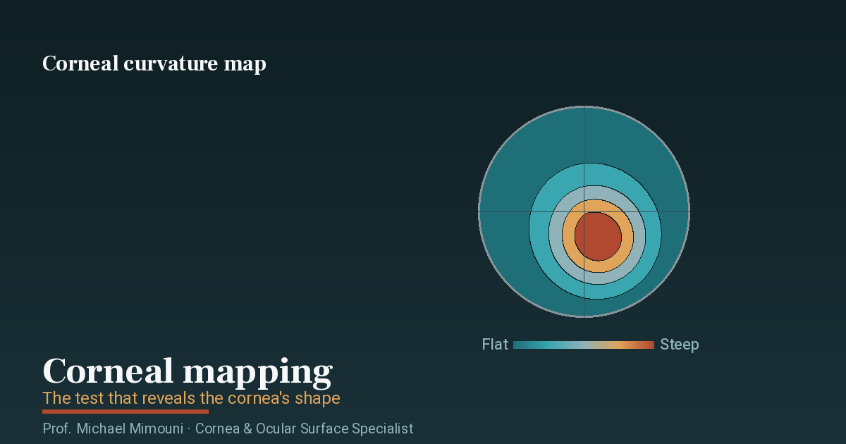
That is why modern formulas take the posterior cornea into account, and it is also why good quality corneal topography is not a formality but the foundation on which the whole decision rests.
How stable is a toric lens
This is a good question, because a toric lens that rotates loses some of its effect. A meta-analysis including 51 studies and 4,863 eyes found a mean absolute rotation of 2.36 degrees. In the best designs, 97 percent and more of eyes stay within 5 degrees.
What happens when they do not: it is commonly estimated that about 3 percent of the effect is lost for every degree of deviation, and that realignment in a short additional procedure is warranted when the deviation exceeds 10 degrees. A note on accuracy: the common rule that at 30 degrees the entire effect is lost is a simplification. Rotation not only reduces the correction but also creates astigmatism on a new axis, so the damage at small rotations may be greater than the simple model predicts.
What the alternatives are
- Corneal relaxing incisions. A Cochrane review compared them directly: about 700 out of 1,000 eyes reached residual astigmatism below half a dioptre with a toric lens, compared with about 500 out of 1,000 with relaxing incisions. A clear advantage to the toric.
- Femtosecond laser incisions. A randomised study in 196 patients found no significant difference between the methods overall — but in the subgroup with 1.5 dioptres and above, the toric was better.
- Placing the main incision on the steep axis. Corrects small amounts only, but costs nothing.
- Additional laser surgery after the cataract operation. Possible, but it turns a plan of one surgery into a plan of two, and therefore it needs to be said before the surgery and not after it.
Halos and glare — the real numbers
This is the part I most want you to read, because it is the part missing from most marketing material.
A meta-analysis of patient reports, eleven studies and 580 patients with a trifocal lens in both eyes:
| Phenomenon | Reported it | Rated it severe | Rated it very bothersome |
|---|---|---|---|
| Halos around lights | 43.9% | 5.4% | 1.4% |
| Glare | 33.6% | 2.9% | 0.8% |
| Starbursts | 30.4% | 3.4% | 2.6% |
The phrasing I use in the clinic: about 4 in every 10 will see halos. About 1 in every 20 will call them severe. About 1 to 3 in every 100 will call them very bothersome.
Two contexts matter for these numbers:
First, an ordinary lens also produces light phenomena. Positive dysphotopsia appears at an early stage in up to about 49 percent of all operated patients, and the phenomenon of a dark shadow at the outer edge persists in about 3 percent at one year — with standard monofocal lenses. The baseline is not zero. Anyone presenting halos as a phenomenon exclusive to premium lenses is misleading you.
Second, regarding the large comparative data. A Cochrane review comparing multifocals with monofocals found a risk ratio of 3.58 for halos and 1.41 for glare, alongside a reduction in spectacle dependence at a risk ratio of 0.63. The direction of these findings is well founded, but the review rests largely on previous-generation bifocal lenses and not on today’s trifocals. I quote it because it is the highest quality evidence that exists, and I qualify it because that is what is accurate.
And a note on EDOF lenses versus trifocals: two meta-analyses examined whether EDOF has an advantage in halos. One found no significant difference, the other found about 32 percent more halos with a trifocal. In other words — the EDOF advantage in this area probably exists but it is modest and has not been demonstrated consistently. Anyone promising you “EDOF without halos” is promising more than the data supports.
Monovision — the option most people have not heard of
Not every route to getting rid of reading glasses goes through an expensive lens. In monovision one eye is set for distance and the other slightly for near, using two ordinary monofocal lenses.
A study of mini-monovision, with the non-dominant eye targeted at minus 0.75 dioptres, 50 patients: 20 percent needed reading glasses in the monovision group compared with 80 percent in the group targeted for distance in both eyes. Light phenomena were reported in only 8 percent. The European guideline phrases it directly: monovision and EDOF lenses suit those who want good vision at intermediate distance, with significantly less dysphotopsia than multifocals.
Who it suits particularly well: anyone who has already succeeded with monovision in contact lenses (that is the best predictor there is), anyone with a contraindication to a diffractive lens, and anyone who prefers contrast and good night vision over full independence in reading. And no less important — it is possible with lenses that are inside the health basket.
Who it does not suit: manifest strabismus, phoria above 8 prism dioptres, previous eye muscle surgery, existing double vision or prism in the glasses, amblyopia with strong dominance, and anisometropia above 1.5 dioptres from childhood. There is also a condition that is easy to miss — monofixation, which appears in about 30 percent of perfectly straight eyes, and which can decompensate precisely when a difference between the eyes is created.
Who a multifocal lens does not suit
Back to the principle: there is no contrast reserve to give up. Everything below follows from it.
Macular disease — macular degeneration, an epiretinal membrane causing image distortion, diabetic macular oedema involving the centre. Two reasons: the near focus of the lens can only be used by the centre of the retina, and a diffractive lens also makes it harder for the physician to view the retina and creates artefacts on OCT — which interferes with follow-up over years.
Here I owe you a qualification. A 2020 review examining multifocal lenses and retinal disease concluded that some of these contraindications are “hypothetical in nature and not evidence-based”. The honest translation: most surgeons will not implant a multifocal lens in an eye with macular disease, and this is based on optical logic and clinical experience more than on randomised trials. That is my practice, and I note it for what it is.
Glaucoma with visual field loss — glaucoma damages contrast sensitivity more than visual acuity, so the losses accumulate. There is also a real follow-up problem: a multifocal lens lowers the mean value on computerised visual field testing and creates artefacts on OCT, in a disease managed over decades on precisely the basis of those tests. Mild, stable glaucoma without central involvement — an EDOF or an enhanced monofocal may possibly be considered. Moderate glaucoma and above — no. Toric and aspheric lenses, by contrast, are entirely suitable in glaucoma.
An irregular cornea or keratoconus — an irregular cornea already produces several blurred images by itself; adding a lens that produces further images doubles the problem. The threshold in clinical use is higher order aberrations below 0.3 microns over a 4 mm diameter. This too is a proposed threshold and not prospectively validated, but it has sound optical reasoning behind it. The only exception with a rationale in an irregular cornea is a small-aperture principle lens, at the price of dimmer vision in low light.
Significant dry eye — and the two reasons are entirely different from one another. The first: dry eye corrupts the measurements on which the surgery is planned, and instability of the tear film translates directly into an error in lens power and in the toric axis. The second: after surgery, a multifocal lens has no spare contrast to waste on an unstable tear film — and cataract surgery itself worsens dryness for months. The accepted approach: severe dry eye disqualifies, and mild to moderate dry eye is treated to stability and re-measured before committing to a premium lens.
A pupil smaller than 2.5 mm, a large angle between the visual axis and the optical axis, a cornea with Fuchs dystrophy even at a mild grade, corneal scars, and any condition expected to cause tilt or decentration of the lens — zonular weakness, pseudoexfoliation, previous vitrectomy surgery.
A further qualification that ought to be stated: the widely used angle thresholds are a convention, not validated values. A 2024 study found no significant correlation between the angle and the outcome or light phenomena in modern diffractive lenses, and challenged the very need for routine measurement. We still measure — but it is worth knowing that this is under debate.
A functionally single eye. Tolerance of a multifocal lens rests largely on the brain using the other eye to suppress the out-of-focus image. Someone with one eye has no partner, and no margin for error either. By the same logic, multifocal implantation in one eye only is tolerated less well than in two.
Occupations dependent on night driving — professional drivers, pilots, night shift work, and also anyone working with a microscope or precision optical equipment, because the fixed working distance of a multifocal lens does not match those instruments. A note on the evidence: I did not find a study quantifying actual night driving performance by lens type. That is to say, it cannot be promised that this is fine and it cannot be proven that it is dangerous. The absence of that datum also deserves to be stated.
And the contraindication hardest to quantify — expectations and personality. Anyone expecting perfect vision at every distance and in every lighting, anyone who was bothered by half a dioptre of error in their glasses before the surgery, anyone who describes their problem in unusually great detail, anyone going through a period of anxiety. And here is the most important admission on this page: there is no validated tool at all for measuring this. The European guideline settles for recommending shared decision-making with the patient, and that is an implicit admission that selecting patients for premium lenses remains a matter of clinical judgement.
If you have previously had laser vision correction
This is a group I see a great deal of, and it deserves its own section — because here the calculation really is different. Someone who had laser vision correction twenty years ago is reaching cataract age today, and rightly expects the same level of result.
Why it is harder — three separate errors that do not cancel each other out:
- The keratometric index error. The devices measure the front of the cornea and infer the total power from it, on an assumption about the ratio between front and back. The laser changed the front and not the back, so the assumption is no longer true.
- The measurement zone error. Standard keratometry samples a ring around the centre, which after myopic treatment lies partly outside the flattened zone — meaning it measures a steeper area than the one the patient actually looks through.
- The effective lens position error. Most formulas use corneal power to predict where the lens will sit. Laser surgery changed corneal power without changing the structure of the front of the eye, so the prediction is biased.
The overall direction: after laser treatment for myopia, an uncorrected calculation selects a power that is too low — and the result is a hyperopic surprise.
What is done in practice: dedicated formulas are used, chief among them the calculator of the American society for cataract and refractive surgery, which aggregates several methods. The European guideline refers to it explicitly. The practical rule is to calculate by several methods and take the median or the mean, and when the methods diverge widely — to lean towards a slightly myopic result, which is easier to live with.
And the honest numbers: in an eye that has not had laser, we land within half a dioptre of target in about 50 to 70 percent of cases, and within a full dioptre in about 80 to 95 percent. After previous laser the figure is lower — in recent series it is around two thirds within half a dioptre. There is also an interesting hint that the difference between LASIK and PRK is meaningful, but it is based on small subgroups and I note it as a hypothesis and not as a fact.
The practical conclusion: in an eye that has had laser, a multifocal lens tolerates an error of a quarter or half a dioptre less well than a monofocal lens does — and it is precisely in that eye that the chance of such an error is greater. That is exactly the combination that produces dissatisfaction. So in a large proportion of cases I recommend, in such an eye, an EDOF, an enhanced monofocal or a toric monofocal — not because advanced technology is “forbidden”, but because in that eye it brings less benefit and more risk. And if you had laser and have suffered from dry eye ever since, that is a further consideration to be dealt with before measuring.
What happens when someone is dissatisfied
This is a section most pages do not write, and I think it is the most important one for the decision.
In a series of patients referred for dissatisfaction with a multifocal lens, the most common cause was not the lens — it was residual refractive error left after the surgery, in 64.5 percent of cases. After it: posterior capsule opacification in 15.8 percent, a large pupil in 14.5 percent, optical aberrations in 11.8 percent, and lens decentration in 9.2 percent. In another series, dry eye was a factor in 35 percent.
The encouraging figure: 84.2 percent of cases were treatable, and lens exchange was required in only 4 percent.
And the figure that must not be hidden: in another series of dissatisfied patients who were actively managed, 45 percent resolved fully, 23 percent partially, and 32 percent remained fully dissatisfied. That is a figure from a referral population and not a general risk, but it is honest evidence that not every dissatisfied patient can be rescued.
What I take from this, and what you should take: most of what goes wrong with a premium lens is not the choice of lens — it is the quality of the measurements, the stability of the ocular surface, and what was or was not said before the surgery. That is why time invested before the surgery is worth more than any technological upgrade.
Capsule opacification — a point that touches on lens choice
Posterior capsule opacification is the most common phenomenon in the years after cataract surgery, and it is treated with YAG laser. It is relevant to this page for two reasons.
First, the lens material matters significantly. A British study of 20,763 eyes found YAG treatment rates within 5 years of 5.8 percent and 8.5 percent in hydrophobic acrylic lenses, compared with 15.2 percent and 19.3 percent in hydrophilic lenses — a threefold gap. A sharp optic edge also contributes, as does a surgical technique in which the capsular opening overlaps the edge of the lens.
Second, the threshold for treatment is lower with a premium lens. A degree of opacification that a patient with a monofocal lens would not have felt at all measurably degrades the image of a multifocal lens — because opacification scatters light, and such a lens has no spare contrast. This direction is consistent in the literature. I will not publish an exact figure here because I was unable to verify one from a primary source, and I prefer to state the direction without giving a certainty I do not have.
One technical point worth knowing: YAG treatment makes a future lens exchange harder and riskier. So in a patient struggling with a multifocal lens where exchange is still on the table, it is not right to perform YAG automatically.
What cannot be promised
These are the things I say out loud before anyone decides:
Not “you will never need glasses.” The honest promise is a reduction in dependence, quantified by lens type — not independence.
Not “you will not see halos.” About 44 percent see them with a bilateral trifocal. This is not a complication but an expected consequence of the design.
Not “the halos will certainly pass.” Neuroadaptation does reduce the symptoms in most people within weeks to months, but it is not guaranteed and there is no way to predict who will adapt.
Not “we will hit the target exactly.” Even in an ordinary eye a not insignificant proportion of eyes land outside half a dioptre, and after previous laser more so. And a premium lens magnifies the significance of a small miss.
Not “one surgery and that is it.” A significant minority of patients will need a further intervention — YAG, a supplementary laser correction, rotation of a toric lens, or rarely an exchange.
Not “night vision will be the same as with an ordinary lens.” Contrast sensitivity is lower in some designs. That is a measured fact.
Not “if you do not like it, we will simply exchange it.” Exchange is possible and usually successful, but it is a second intraocular surgery with risks of its own, it is harder after YAG, and the refractive predictability of the replacement lens is lower.
Not “a good lens will compensate for another disease in the eye.” No lens improves the retina, the optic nerve or an irregular cornea. In such situations a premium lens can only make the optical side worse.
And not “we know how to predict who will be satisfied.” There is no validated tool for that. That is probably the most honest sentence that can be written on a page like this.
How I approach it
The order I work by is this: first to check what the eye can do — retina, optic nerve, cornea, ocular surface. Then to establish what the patient actually needs: what he does during the day, whether he drives at night, whether he reads a great deal, what would bother him more — reading glasses or halos. And only after those two, to talk about a lens.
A patient whose eye is not suited to a multifocal lens and who receives one has paid more and received less. That is the outcome I try hardest to prevent — more than achieving the most impressive result in the suitable case.
If you are undecided and want a personal assessment before you decide, you are welcome to contact me. And if you are also looking into the financial side, I set it out on the cataract surgery cost page.
Frequently asked questions
Which is the best lens for cataract surgery?
There is no single best lens. A monofocal lens gives the best distance acuity and the best contrast sensitivity, but requires reading glasses. A multifocal lens gives the greatest independence from glasses, but at the cost of halos around lights and lower contrast sensitivity. The right choice follows from the state of the specific eye and from the patient's own order of priorities, not from a general ranking.
How many people see halos after a multifocal lens?
In a review pooling eleven studies and 580 patients with a trifocal lens in both eyes, about 44 percent reported halos, about 34 percent glare and about 30 percent starbursts. About 5 percent rated the phenomenon as severe and about 1 to 3 percent as very bothersome. Halos are not a complication — they are an expected optical consequence of the lens design.
When is a toric lens needed?
The guidelines of the American Academy of Ophthalmology and of the European society agree that a toric lens should be considered from 1.0 dioptre of corneal astigmatism upwards, and that the clearest benefit is above 2.0 dioptres. Below that threshold there is genuine disagreement between surgeons, and correction is also possible with relaxing incisions or by placing the main incision on the steep axis.
Can a multifocal lens be implanted after laser vision correction?
It can, but this is one of the situations that demands the greatest caution. A cornea after laser already has a multifocal character, lens power calculation is less accurate, and these patients usually have high expectations. In an eye that has not had laser, we land within half a dioptre of target in about 50 to 70 percent of cases; after previous laser the figure is lower. In a large proportion of cases an extended depth of focus lens or an enhanced monofocal is the more appropriate choice.
Who is a multifocal lens not suitable for?
Mainly anyone with macular disease, glaucoma with visual field loss, an irregular cornea or keratoconus, significant untreated dry eye, a very small pupil, or a functionally single eye. The reason is common to all of them: a multifocal lens splits the light between two or three foci, and any eye that has already lost contrast has no surplus to give up.
What is the most common reason for dissatisfaction after a premium lens?
Not the lens itself. In series of patients referred for dissatisfaction, the most common cause was residual refractive error left after the surgery — 57 to 64.5 percent of cases — followed by dry eye and posterior capsule opacification. The good news is that most of these factors are correctable.